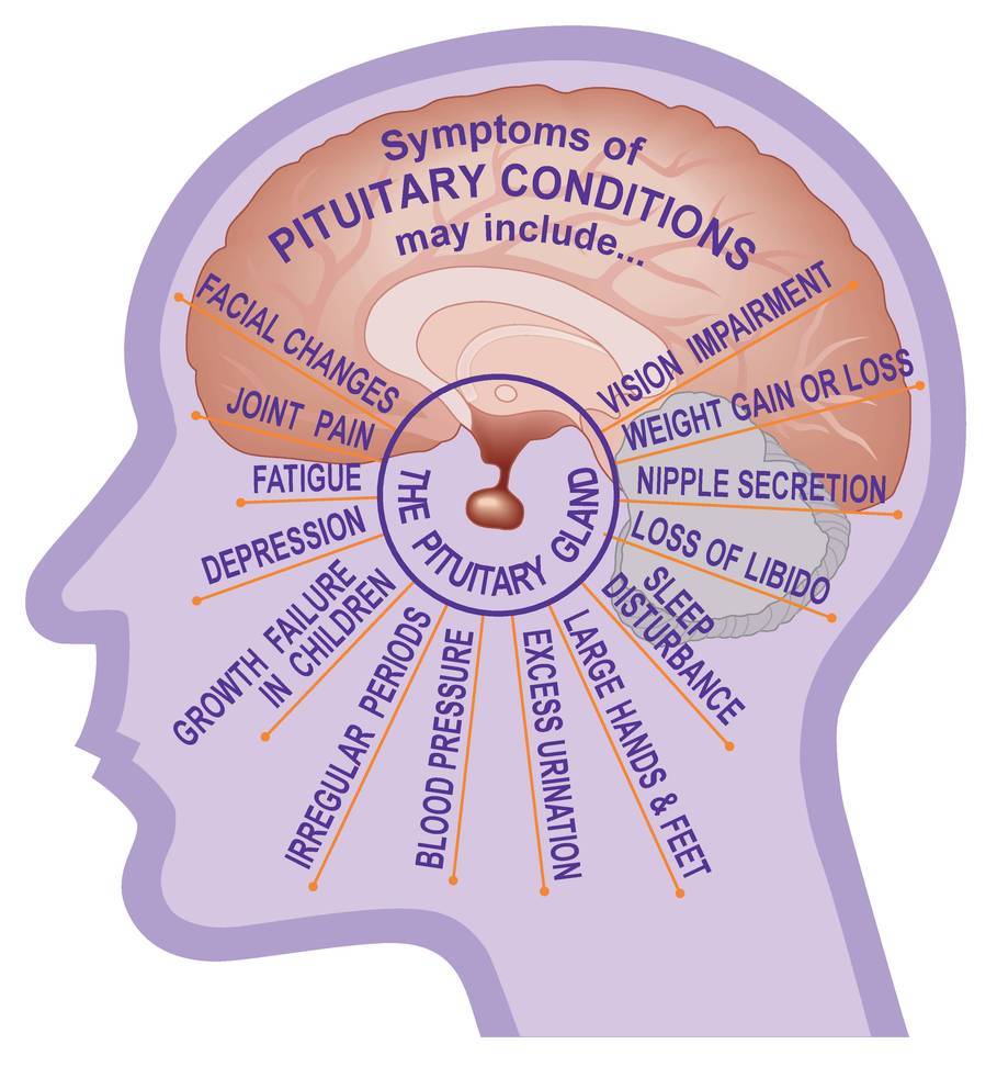
Symptoms
Pituitary tumours can cause headaches and lead to vision disorders. There are several types of pituitary tumours, the common types being adenomas, craniopharyngiomas and Rathke’s cleft cysts. Most pituitary adenomas are non-cancerous, slow-growing and often curable. Tiny tumours or cysts are often detected in MRI scans. Most of these are harmless; they never grow further or cause any symptoms.
Functioning pituitary tumours can produce high levels of hormones that interfere with other bodily processes. The tumours function in accord with their cell of origin and are named for the particular hormone they produce. For instance, prolactinoma originates in cells that produce prolactin.
Non-functioning adenomas do not produce hormones. They grow till their size and mass start causing symptoms like headaches, vision loss, nausea, vomiting and fatigue. Such tumours may constrict the pituitary gland, which leads to decrease in hormone production (hypopituitarism) till it completely stops.
Pituitary tumours less than 10mm are classified as microadenomas and larger ones as macroadenomas. Large adenomas can apply pressure on the optical nerves and infest the cavernous sinuses, which are home to the carotid arteries and nerves governing eye movement.
Craniopharyngiomas and Rathke’s cleft cysts are closely related to pituitary adenomas. They grow from the remnant embryonic cells in the pituitary gland. Craniopharyngiomas usually originate in the pituitary stalk and grow upward into the third ventricle. Symptoms are similar to pituitary adenomas.

Diagnosis
Any diagnosis begins with the doctor obtaining the patient’s personal and family medical history followed by a physical examination. For Pituitary tumours, physicians also perform a neurological exam to check overall mental condition including memory, muscle strength, cranial nerve function, coordination and response to pain. There may also be one or more of the following tests:
- Magnetic Resonance Imaging (MRI) scan
- Endocrine evaluation
- A visual field acuity test
- Petrosal sinus sampling
Treatments
Treatment options depend on the type, size, stage and location of the tumour besides age and general health of the patient. The goal of treatment is to eradicate the tumour or control its growth. Hormone levels are corrected with medications. Main choices are surgery, radiotherapy, chemotherapy and endocrine therapy either alone or in combination.
A neurosurgeon, ENT surgeon, endocrinologist and radiation oncologist work as a team to determine the optimal treatment for a particular patient. Hence, it is important to receive treatment at a medical centre, which offers all treatment options.
Surgery
Surgical elimination of a pituitary tumour can involve minimally invasive endoscopic transsphenoidal surgery, conventional microscopic transsphenoidal surgery or traditional craniotomy. The choice varies from patient to patient depending on the type, size and location of the tumour.
Surgery is the main mode of treatment for tumours producing growth hormones and
Adrenocorticotropic Hormone (ACTH) to manage the endocrine system. In case of tumours located near vital tissues and nerves, the neurosurgeon may remove them only partially, which can still alleviate symptoms and stabilize hormone levels. Radiation therapy then targets the remaining part of the tumour.